
Nevus lipomatosus cutaneous superficialis - An odd site
*Corresponding author: Yogindher Singh, Department of Dermatology, Venereology, Leprosy, Sri Venkateshwaraa Medical College, Puducherry, India. yogindher@gmail.com
-
Received: ,
Accepted: ,
How to cite this article: Prasanna A, Singh Y. Nevus lipomatosus cutaneous superficialis - An odd site. Karnataka Med J. 2024;47:43-4 doi: 10.25259/KMJ_1_2024
Dear Sir,
Nevus lipomatosus cutaneous superficialis (NLCS) is an uncommon idiopathic hamartomatous condition characterised by ectopic collections of mature adipocytes in reticular dermis.[1] First notified by Hoffmann and Zurhelle in 1921.[2] Mainly classified into two – CLASSICAL and SOLITARY based on morphology, of which the classical type usually seen over the pelvic girdle, lumbar, buttocks, and upper thighs.[1-3] Herein, we report a case of classical-type NLCS presenting over the back, an unusual site.
An otherwise normal 16-year-old girl came to our outpatient department with asymptomatic numerous elevated skin lesions over her right upper back for the past eight years, which gradually progressed in size.
Dermatological examination revealed multiple skin-coloured and hyperpigmented papules and cerebriform nodules coalescing to form plaques with segmental distribution over the right shoulder and upper back [Figure 1a]. There were no features suggestive of neurological impairment and no café-au-lait macules found anywhere in the body.
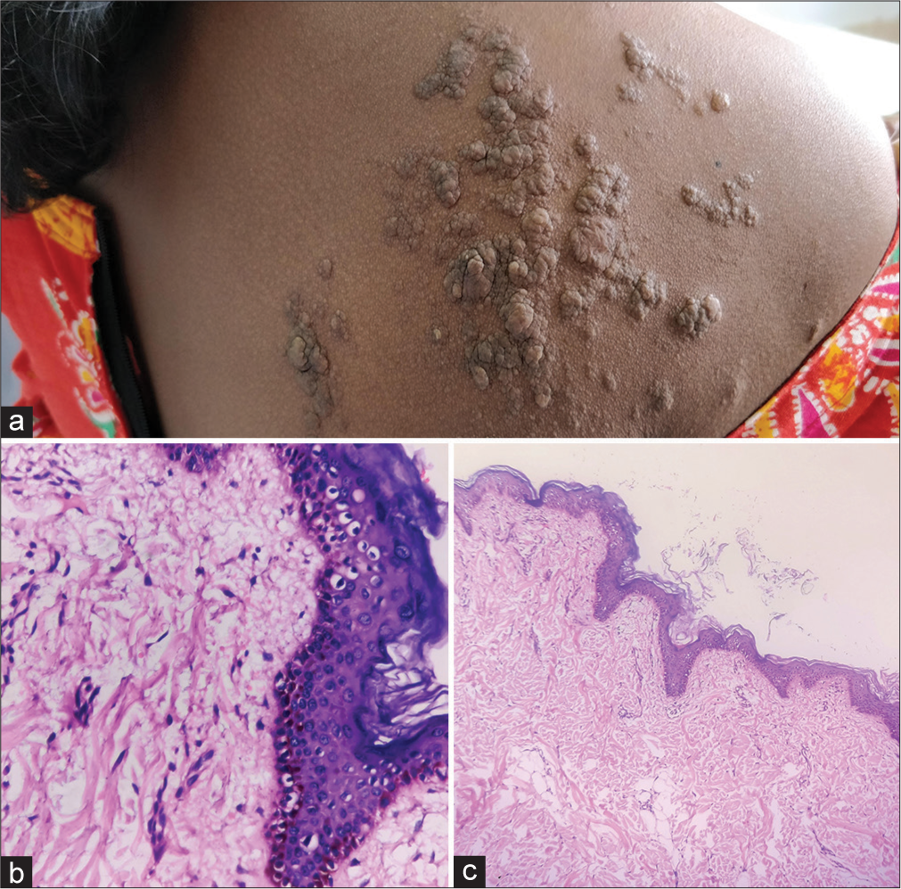
- (a) Multiple skin coloured and few hyperpigmented papules and cerebriform nodules coalescing to form plaques seen in segmental distribution. (b) Adipose tissue island is seen almost reaching papillary dermis. (c) Hyperkeratosis, acanthosis, papillomatosis along with keratotic plugging. Dermis showed admixture of collagen and adipose tissue islands.
Routine investigations were normal, followed by which 4-mm lesional punch biopsy was taken. Histopathological analysis showed keratotic plugging, minor hyperkeratosis, acanthosis, papillomatosis, and increased melanin pigment. Adipose tissue islands and collagen were mixed together in the deeper tissue, almost reaching the papillary dermis [Figure 1b and c].
NLCS is an idiopathic, benign, hamartomatous nevoid anomaly attributed to the presence of mature ectopic fat in the dermis.[1-5] Morphologically, categorised into uncommon classical (multiple) types and common solitary types.[3-5] Classical type presents as asymptomatic, soft, skin coloured to yellow papulo-nodules, which may coalesce to form a plaque with a smooth, wrinkled and cerebriform surface. It follows zosteriform distribution and commonly occurs over the pelvic girdle, buttocks, lumbo-sacral region and thigh and presents after birth or within a few decades of life.[1-5] On the contrary, solitary type presents as a single papule or nodule with no site predilection and appears commonly during the third to fourth decades of life.[4,5] NLCS has neither sex preference nor associated organ involvement or malignant transformation.[4] However, the coexistence of pigmentary conditions such as café-au-lait spots, leucoderma, capillary haemangioma, trichofolliculoma, hypertrichosis and comedo-like lesions has been reported.[2-4]
There are no clear-cut aetiology and pathogenesis for NLCS. Degenerative changes in the dermal collagen and elastic tissue resulting in the deposition of adipose tissue in the dermis are postulated by Hoffmann and Zurhelle. Another theory is that adipose metaplasia in the dermal connective tissue is linked to NLCS.[1,4] Electron microscopy may reveal lipocytes in the presence around capillaries which denotes hamartomatous origin from the pericytes. [3] Histopathological features suggest NLCS-ectopic mature adipocytes in the perivascular region of the dermis.[1,4]
Clinical differential diagnosis includes fibroepithelial polyp, lymphangioma, neurofibromatosis and acrochordon, which can be distinguished histologically.[2-4]
Histopathological differential diagnosis includes melanocytic nevi, glotz’s syndrome, acrochordon,[1,3,4] by the presence of ectopic mature adipocytes in the dermis, in our case almost reaching the papillary dermis.
As NLCS is a benign condition treatment that is usually unnecessary, if required for cosmetic purposes, a simple surgical excision can be done, which appears to be curative.
In conclusion, we are reporting this case for its rarity and, in our case, for its odd site (upper back). Hence, early diagnosis and prompt treatment aids in cosmetic concerns and improves the quality of life of the patient.
Ethical approval
Institutional review board approval is not required.
Declaration of patient consent
The authors certify that they have obtained all appropriate patient consent.
Conflicts of interest
There are no conflicts of interest.
Use of artificial intelligence (AI)-assisted technology for manuscript preparation
The authors confirm that there was no use of artificial intelligence (AI)-assisted technology for assisting in the writing or editing of the manuscript and no images were manipulated using AI.
Financial support and sponsorship
Nil.
References
- Nevus lipomatosis cutaneous superficialis-a clinicopathologic study of the solitary type. Med J Armed Forces India. 2016;72:67-70.
- [CrossRef] [PubMed] [Google Scholar]
- About a nevus lipomatosus cutaneous superficialis of the left gluteal region. Arch Dermatol Syphilol. 1921;130:327-33.
- [CrossRef] [Google Scholar]
- Nevus lipomatosus cutaneus superficialis associated with nevus sebaceous of Jadassohn. Indian J Dermatol Venereol Leprol. 2014;80:194.
- [CrossRef] [PubMed] [Google Scholar]
- Huge nevus lipomatosus cutaneous superficialis on back: An unusual presentation. Indian J Dermatol. 2015;60:296-7.
- [CrossRef] [PubMed] [Google Scholar]
- Nevus lipomatosus cutaneous superficialis with unusual presentation over the nipple. Indian J Dermatol. 2017;62:429-31.
- [CrossRef] [PubMed] [Google Scholar]


